Explore our diagnostic imaging services
A nuclear medicine exam used to assess blood flow to the heart muscle at rest and during stress.
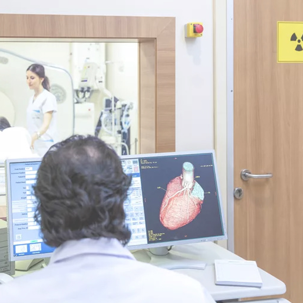
Myocardial Perfusion Imaging (MPI) is a nuclear medicine exam used to evaluate blood flow to the heart muscle and identify areas with reduced circulation. The test helps determine whether blockages may be present in the coronary arteries by comparing blood flow during rest and during stress.
MPI provides important functional information that supports diagnosis and management of heart disease.
Myocardial perfusion imaging is performed in two parts—one at rest and one under stress—often on separate days.
During the rest portion, a small amount of radiopharmaceutical is injected into a vein in your arm. After a brief waiting period of approximately 30–60 minutes, images of your heart are obtained using a gamma camera. Imaging typically takes about 20 minutes.
During the stress portion, blood flow to the heart is increased either through exercise on a treadmill or, if exercise is not possible, with a medication such as Persantine®. A small amount of radiopharmaceutical is injected at peak stress, followed by a waiting period and imaging similar to the resting portion.
You may be asked to bring a list of your medications, as certain medications may need to be adjusted prior to the stress portion of the exam.
Evaluate blood flow to the heart muscle
Detect reduced circulation or coronary artery blockages
Compare heart perfusion at rest and during stress
Support diagnosis and management of heart disease
The amount of radiation used is minimal, and MPI has a long history of safe and effective clinical use.
Physicians: These can be taken to any licensed facility providing healthcare services including hospitals and IHFs. Please click the button below to download an editable .pdf copy of our requisition and send it with your clients before their appointment.